Ads
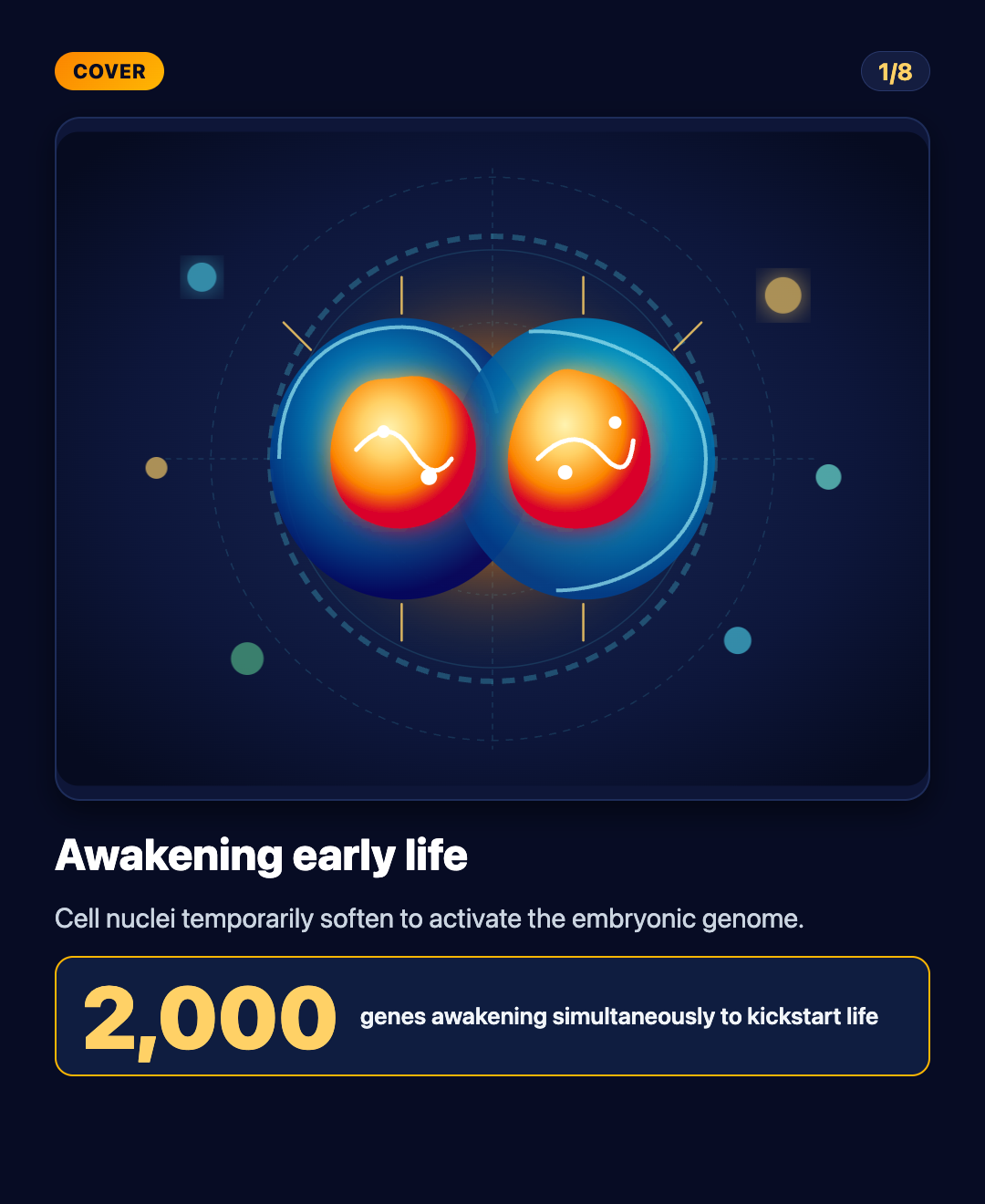
Embryonic cell nuclei temporarily soften to awaken the machinery of life
3 ngày trước
00Imagine walking into a densely packed library where every door is locked from the inside and thousands of irreplaceable blueprints are trapped under thick ice. For this building to function, someone must enter and open every page simultaneously. That is precisely the challenge that every living creature on Earth faces in the earliest moments of existence.
On November eleventh, twenty twenty-six, in a study published in the journal Nature, a research team led by Professor Kei Miyamoto at Kyushu University and Associate Professor Yuta Shimamoto at the National Institute of Genetics in Japan unlocked this long-standing mystery. They discovered that the nucleus of a two-cell embryo automatically softens into a gelatin-like state, temporarily dismantling its protective scaffolding to awaken the dormant genome.
Note – Cell nucleus: The spherical command center of a cell, acting like a secure vault holding all the DNA blueprints of life.
WHAT WAS THE PROBLEM TO BEGIN WITH
After sperm fertilizes an egg, the resulting single cell divides into a two-cell embryo. At this juncture, the embryo faces an existential countdown: maternal nutrients and regulatory proteins inherited from the egg are rapidly depleted. The embryo must take control and activate its own genes, a developmental milestone known as zygotic gene activation.
Note – Major zygotic gene activation: The critical developmental milestone where the embryo turns on its own genetic switch, awakening around two thousand genes to begin constructing an organism.
For decades, biologists believed this awakening was governed strictly by chemical keys. The prevailing model assumed that specialized regulatory proteins arrived, docked onto DNA strands, and unlocked specific genes. Yet a fundamental physical paradox remained unanswered: how could thousands of tightly wound genes within a cramped, microscopic nucleus all burst open within a brief window without major physical reorganization?
WHAT JUST CHANGED COMPARED TO OLD BELIEFS
The new research overturns conventional wisdom by proving that mechanical physical forces are the primary drivers. Instead of maintaining a rigid shell to shield genetic material as mature adult cells do, the nucleus of a two-cell embryo undergoes a striking, temporary transformation, becoming exceptionally soft and pliable.
Scientists found that the internal structural scaffolding beneath the nuclear envelope is deliberately disassembled. Much like removing steel crossbars from a building frame to allow its walls to flex freely, this nuclear softening gives internal chromatin fibers space to loosen, expand, and interact smoothly with biological transcription machinery.
Note – Nuclear envelope: The double-layered membrane encasing the nucleus, serving as a protective barrier shielding DNA from external physical stress.
HOW SCIENTISTS FIGURED THIS OUT
The team in Japan used ultra-fine glass micropipettes to directly probe the nuclei of living mouse embryos and measure their elastic mechanical properties. They were astonished to find that nuclear stiffness at the two-cell stage dropped abruptly by more than fifty percent compared to fertilized eggs, turning as squishy as loose gelatin.
At the molecular level, they traced this softening to a structural protein named Lamin B1. Under normal conditions, Lamin B1 polymerizes into a dense, load-bearing fibrous mesh directly beneath the nuclear envelope. During the two-cell stage, the embryo mobilizes a molecular recycling machinery named LC3 to selectively dismantle and clear away Lamin B1.
Note – Lamin B1: A fibrous structural protein forming the primary load-bearing scaffold of the nuclear envelope, similar to steel reinforcement in concrete.
Note – LC3 protein: A cellular housekeeping factor that acts like a waste management team, tagging and clearing structural proteins to reorganize the cell.
To confirm that softening is essential for life rather than an incidental artifact, the researchers artificially tethered nuclei to maintain their rigidity. The outcome was stark: when nuclei were artificially kept stiff, DNA failed to expand, approximately two thousand embryonic genes remained dormant, and embryo development ground to an immediate halt. Only when the nucleus was allowed to soften could embryonic life proceed.
WHAT THIS DISCOVERY MEANS FOR HUMANITY
This discovery provides a transformative perspective on the physical origin of life, forging a direct bridge between classical mechanical physics and molecular genetics. It shows that life is not merely a symphony of chemical reactions, but a synchronized ballet of biophysical forces, surface tensions, and microscopic elasticity.
Looking forward, these findings hold significant promise for assisted reproductive technology and in vitro fertilization. Currently, fertility specialists evaluate embryo quality primarily through optical visual inspection, an assessment method prone to uncertainty. Measuring the mechanical compliance of embryonic nuclei could become an objective physical biomarker, helping clinicians identify the healthiest embryos and improving success rates for families facing infertility.
Note – In vitro fertilization: An assisted reproductive procedure in which eggs and sperm are combined outside the body in a laboratory before a healthy embryo is transferred to the uterus.
Furthermore, the study provides valuable insights into oncology. Metastatic cancer cells frequently soften their nuclei to squeeze through narrow capillaries and invade distant tissues, echoing the cellular behavior observed in early embryos. Deciphering how the Lamin B1 scaffold is regulated could help oncologists devise therapies to prevent cancer cells from softening and spreading.
Nevertheless, translating these laboratory insights into clinical care will require years of research. This study was performed on animal models. Human embryos are delicate and governed by strict ethical considerations, precluding direct invasive micropipette probing. The next task for the scientific community is to engineer gentle, non-invasive optical tools that can evaluate nuclear compliance without compromising developing life.
SOURCES
Nature – Transient nuclear softening for zygotic gene activation in mice – https://doi.org/10.1038/s41586-026-11240-w
Kyushu University – Department of Stem Cell Biology – https://www.kyushu-u.ac.jp
National Institute of Genetics – Division of Cell Biophysics – https://www.nig.ac.jp
0 comments
Ads
Related Posts
Ads